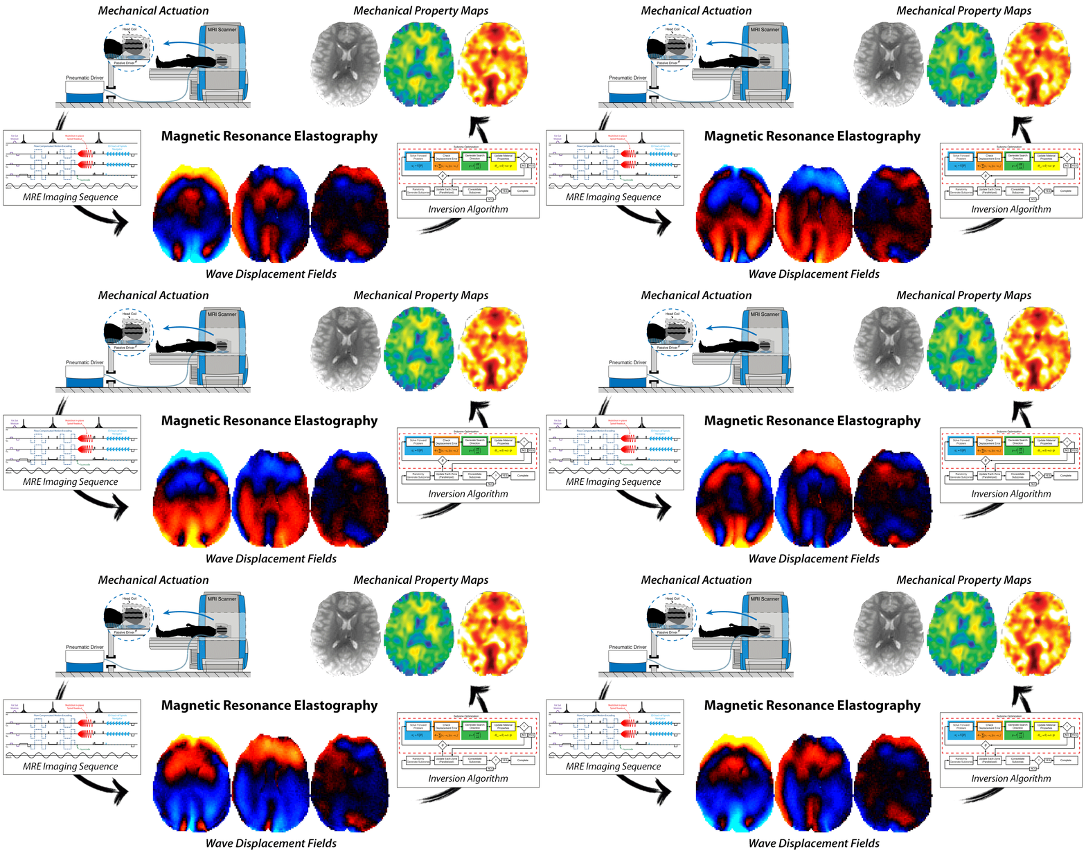
In the Mechanical Neuroimaging Lab, we work to mechanically mapping the brain with magnetic resonance elastography to characterize the structure, function, and health of neural tissue.
We perform research at the Center for Biomedical Imaging at the University of Delaware on the Siemens 3T Prisma scanner and Bruker 9.4T animal scanner. We enjoy strong collaborations with researchers throughout UD Engineering, Health Sciences, and Psychology, and with local partner hospitals including Nemours/A.I. DuPont Hospital for Children.
Magnetic Resonance Elastography
We develop high-resolution methods for MRE to improve the ability to accurately and reliably map the mechanical properties of the human brain in vivo. These developments include:
MRE pulse sequences for improved resolution and speed
Image reconstruction algorithms to improve data quality and limit artifacts
Hardware to actuate tissue in different configurations
Pre-clinical methods for mechanistic studies of brain mechanics
We couple our approaches with the nonlinear inversion (NLI) algorithm, developed by our colleagues at Dartmouth College, to provide high-quality maps of brain tissue viscoelastic properties.
We share our methods widely with researchers and clinicians interested in incorporating MRE in their scan protocols. If interested in collaborating and obtaining sequences for brain MRE, please contact Dr. Curtis Johnson (clj@udel.edu).
Aging Brain Mechanics
We are using MRE to study how brain mechanical properties change with aging, how they reflect cognitive decline, and are affected by lifestyle factors like exercise. Ongoing clinical projects examine mild cognitive impairment and Alzheimer’s disease.
Pediatric Brain MRE
Our group is developing new, fast MRE methods to study pediatric populations to better understand brain maturation. We have active projects in typical and atypical development, including autism and cerebral palsy, and in characterizing pediatric brain tumors.
Traumatic Brain Injury
Mechanical properties of the brain are critical for predicting, modeling, and assessing traumatic brain injuries (TBI). We are using MRE to build models of TBI, determine how the brain moves and deforms on impact, and how brain health changes due to repeated head impacts in hockey players.


